Image

LOS
The Laser, Optics & Spectroscopies (LOS) platform at the University of Namur offers unique expertise in the study of the rotational, vibrational, electronic and scattering properties of matter, explored via linear and non-linear optical processes generated by dedicated lasers and optical sources.
It enables the analysis of gaseous traces, molecular films at interfaces, as well as 2D and 3D nano-systems.
To complement its experimental skills, the platform develops analytical and computational models to interpret photon-matter spectroscopic responses.
The LOS platform applies advanced optical measurements to characterize matter, light and their interactions, as part of fundamental or applied research.
Optical phenomena are used to study matter in the gaseous and solid phases, with targeted themes such as:
The LOS platform boasts an exceptional array of instruments, including:
The platform is open to internal and external researchers.
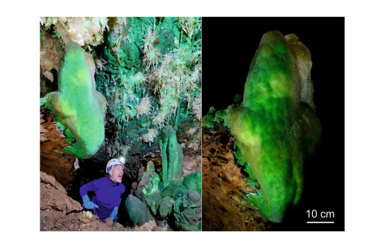
Well hidden from passersby, caves nevertheless conceal particularly aesthetic secrets. For the past four years, Martin Vlieghe has been pursuing a PhD in geology at UNamur. He is exploring the origin of the surprisingly varied colours of certain concretions nestled in the heart of Belgian and French caves. Together with Prof. Johan Yans and Gaëtan Rochez, he samples, observes, and analyses these magnificent objects with the aim of uncovering the mysteries they conceal.
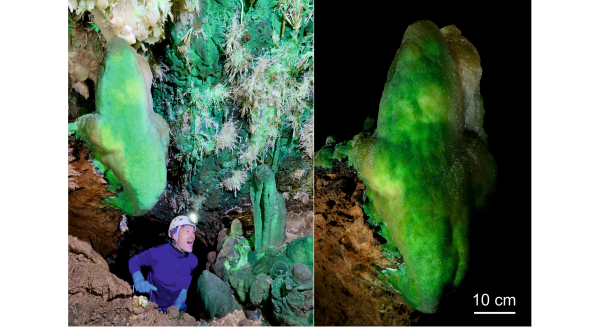
Photo: Green speleothems in the Aven du Mont Marcou (Hérault, France) © Stéphane Pire, Gaëtan Rochez (UNamur)
Speleothems, for instance stalactites and stalagmites, are commonly composed of calcite or aragonite (CaCO3). This mineral compound comes directly from the rock in which the cave was formed and naturally has a white to brownish colour. However, speleothems can sometimes exhibit unique and unusual colours. From yellow to black, blue, red, green, and even purple, there is something for everyone!
Such a diversity of colours reflects the many possible causes: mineralogical, chemical, biological, or even physical. A speleothem, like any natural formation, is never perfectly pure. Their deposition process, through the precipitation of calcium carbonate dissolved in water, is necessarily accompanied by the deposition of numerous impurities carried along with the water circulating underground. Even if these impurities are sometimes too low in concentration or simply uncoloured, they can still have a visible impact on the colour.
The formation of speleothems is very often linked to impurities dissolved in groundwater. Therefore, studying coloured speleothems provides valuable information about potential contamination of surface water with heavy metals or other harmful organic compounds, which in some cases may be consumed by residents. It is therefore a simple and direct way to identify areas with potentially contaminated water and to determine whether this contamination poses an environmental or health risk.
This is the objective of Martin Vlieghe's thesis: to apply a range of cutting-edge analytical techniques to samples of these speleothems to determine these causes and propose an explanation for the origin of the colouring elements.
Here are a few examples.
An initial project explored the green speleothems of the Aven du Marcou (see photo above). Located in the Hérault department of France, this chasm is well known in the area for its series of impressive shafts, the largest of which is over 100 meters deep. It also has a tiny chamber hidden at the top of a steep wall, which houses an impressive concentration of deep green speleothems. After all the effort of descending and climbing ropes to progress through this very vertical cave, what a wonderful reward to discover this true underground gem! Once the initial wonder has passed, it's time to get to work! We observe, describe, interpret, and collect a few green fragments from the ground, while respecting the integrity of the site as much as possible. Back in Belgium, it's time to move on to the analyses.
Careful observation of the recovered fragments quickly reveals the presence of green minerals in the outer part of the speleothems, which are easily associated with the green colour observed. These minerals, which are deposited in platelets parallel to the white aragonite (CaCO₃), turn out to be nepouite crystals, a nickel phyllosilicate ((Ni,Mg)₃Si₂O₅(OH)₄) usually found in marine volcanic rocks.

The discovery is all the more surprising given that there are no nickel deposits in the vicinity of the cave! Further study of the composition of the nepouite reveals that they contain a high concentration of zinc, which is also very unusual and suggests that they are in fact quite different from those commonly mined in volcanic deposits. Finally, this mystery was solved by a thorough examination of the rock outcrops in the immediate vicinity of the cave. Just above the cave are siliceous deposits particularly rich in pyrite, an iron sulphide commonly found in this type of settingst. Analysis of these sulphides reveals high concentrations of nickel, which is also found in the natural water source closest to the cave.
The result of this "investigation" and final explanation: nepouite was able to settle underground through the dissolution of various chemical elements contained in the pyrite of the overlying rocks, which were transported into the cave by surface water and were able to crystallize on site.
The Malaval cave is very different from the Aven du Marcou. Located in Lozère (France), it extends largely along a high underground river that winds beneath the Cévennes massif. At the bend of a meander, one can find magnificent blue speleothems.
As in the Aven du Marcou, the coloured speleothems are found only in two specific locations in the cave and nowhere else, suggesting that the origin of the chromophore elements is probably very localized.
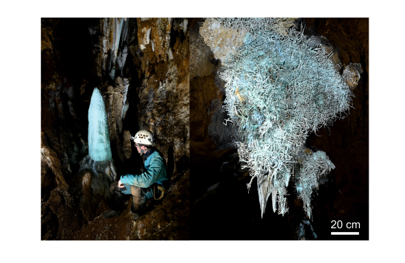
Photos - Left: Blue stalagmite in Malaval Cave. Right: Cluster of blue aragonites in Malaval Cave © Gaëtan Rochez (UNamur)
Once again, a few fragments were collected, including a large bluish stalactite found broken on the cave floor. A series of microscopic observations and mineralogical and geochemical analyses were carried out. The first striking finding was that several blue fragments contained no minerals other than aragonite, suggesting that, unlike the green ones from Marcou, it was the aragonite itself that was coloured by the presence of metallic elements. After examining the analyses, three of these elements stood out: copper, commonly cited as the cause of blue colouring in aragonite, as well as zinc and lead.
While copper appears to be the main cause of the blue colouration, zinc and lead also play a role here.
Zinc is largely present in the form of deep blue amorphous phases, which are only found in some of the blue fragments studied. The presence of these phases, linked to the oxidation of nearby zinc-rich deposits, generates variations in the blue colour at the microscopic level, as revealed by optical microspectrophotometry.

Lead also has a marked colouring power, producing green to blue hues, but statistical analysis of coloured and uncoloured areas shows that these colours only appear in the absence of zinc, which seems to inhibit lead-induced colouring. This study clearly demonstrates that, even if a problem seems easy to explain at first glance, it can sometimes hide unexpected subtleties that need to be explored in greater depth in order to uncover all its secrets.
The Cigalère Cave is one of a kind. Not only does it contain impressive quantities of gypsum, a calcium sulphate found in certain caves, but this gypsum also displays a wide variety of colours rarely seen in nature. Because of this rarity, the cave is particularly well protected, to the point that we were not allowed to collect any fragments from inside it.
This study was therefore the ideal opportunity to test the Geology Department's new acquisition: a portable X-ray fluorescence spectrometer (pXRF), which allows rapid, in situ, and above all completely non-destructive analysis of coloured speleothems.
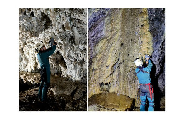
Photos - pXRF analysis of a blue stalactite core (left) and a yellow flowstone (right) in the Cigalère Cave © Stéphane Pire (UNamur)
A total of five sites of interest were selected in the Cigalère for the diversity of colours found there. The pXRF revealed the presence of several metals.
At Cascade Noire, for example, a high concentration of iron in the form of oxides and sulphates was detected, which are responsible for the black and orange colouring of the gypsum, respectively.

Black is also found in the Chapelle de Donnea, but contrary to what one might think, no iron has been detected. Here, it is manganese in the form of oxides that is responsible for the colouration. This observation is interesting because it clearly demonstrates that black colouration in gypsum, two phenomena that appear similar at first glance, can have very different causes, hence the importance of being able to carry out analyses directly in the field.
A little further downstream, blue dominates along the main gallery, and analyses have shown strong similarities with the blue speleothems of Malaval, with a marked influence of copper and potentially zinc.
All this highlights that, despite certain limitations of the device, this type of non-destructive analysis method is a very valuable tool for studying rare, fragile, precious, or protected objects, of which the Cigalère cave is an excellent example!
Martin Vlieghe's doctoral thesis on "The origin(s) of colored speleothems in caves," supervised by Professor Johan Yans and in collaboration with Gaëtan Rochez, began in February 2022. All three researchers are members of the Faculty of Sciences, Department of Geology at UNamur and the ILEE Research Institute.
ILEE (Institute of Life, Earth and Environment) is directly involved in issues related to the study and preservation of the environment, to which this subject is directly linked.
The various analyses were carried out with the support of UNamur's technological platforms:
Some analyses were carried out in partnership with KUL, MRScNB and UMontpellier, and access to the caves was provided by the Association Mont Marcou, the Malaval Association and the Association de Recherche souterraine du Haut Lez.
This thesis was originally funded by the ILEE institute and institutional funds from UNamur, and by an Aspirant F.R.S. - FNRS grant (FC 50205) since October 2023.
It is also closely linked to the new research partnership supported by the RELIEF network (Réseau d’Échanges et de Liaisons entre Institutions d’Enseignement supérieur Francophones), the ILEE research institute at UNamur, and EDYTEM (Environnements, Dynamiques et Territoires de Montagne, Université Savoie Mont Blanc). Mobility programs between these entities will strengthen a common research area: the study of the critical zone, the most superficial zone of the Earth, where rocks, water, air, and living organisms interact. The perspective is to develop other transdisciplinary research areas and potential teaching projects in the field of environmental sciences and sustainable development.
Studying geology means developing a solid foundation in physics, chemistry, and biology in order to understand the Earth, from its internal dynamics to surface processes and their interactions with our environment and human activities.
Thanks to their interdisciplinary training, geologists are ideally positioned to perform a variety of roles that require a rigorous scientific approach to solving complex problems (research and development, project management, consulting, and education).

In a new study published in February 2023 in the Journal of Luminescence, an international team of scientists led by Sébastien Mouchet of UNamur has revealed previously unknown fluorescent properties of the transparent wings of certain insects. This research demonstrates that valuable information can be obtained using state-of-the-art optical characterization techniques. Here’s how it works.

In the natural world, many animal species exhibit fluorescence, meaning they emit visible light under ultraviolet light. In general, the physical, chemical, or biological properties underlying fluorescence in these species are poorly understood. For example, the fluorescence of the transparent wings of the more than 3,000 species of cicadas had never been reported in the scientific literature until this year.
In a new study published in February 2023 in the Journal of Luminescence, an international research team led by Sébastien Mouchet, a researcher in the Department of Physics and a member of the NISM (Namur Institute of Structured Matter) and ILEE (Institute of Life, Earth and Environment) at UNamur, has revealed the previously unknown fluorescent properties of the transparent wings of certain insects, including cicadas found in southern France and the gas-colored sphinx moth, a lepidopteran found in Belgium, among other places.
This study suggests that fluorescence light emission is more common in the transparent wings of insects than previously thought. Everything points to the presence of resilin within the wings as the source of this fluorescence. This protein is known to contribute to the flexibility of the wings.
Contrary to previous beliefs, the fluorescence observed in these insects may be an unintended consequence of resilin’s presence in the wings for mechanical purposes and may not play a specific role in the insect’s visual communication, whether for courtship or territorial defense.
The optical characterization we conducted also allowed us to gain a better understanding of the wing’s structure. The techniques we used are rarely employed in studies of naturally fluorescent organisms. Our research has thus demonstrated that valuable insights can be obtained using these techniques.
The fundamental study of fluorescence in living organisms is not only crucial from a zoological perspective. The discovery of green fluorescent protein (GFP) in the tissues of a species of jellyfish revolutionized the fields of genetics and fluorescence microscopy. This discovery was honored with the Nobel Prize in Chemistry in 2008.
At UNamur, these studies were conducted using equipment from the LOS (Lasers, Optics, and Spectroscopies) technology platform.
The platform offers unique expertise in characterizing the optical and electronic properties of matter using linear and nonlinear optical measurements, performed in particular with lasers within the context of basic or applied research. The platform enables the analysis of structured systems at the nanoscale in one, two, or three dimensions, as well as molecular films at interfaces or trace gases. In addition to its experimental capabilities, the platform develops analytical and numerical models to interpret the measured spectroscopic responses.
This feature is commonly used in our daily lives. Here are a few examples:
Fluorescence is also used in so-called “black light,” which is light emitted by a source composed primarily of near-ultraviolet rays. This light highlights white and fluorescent objects to create a special atmosphere, verify that a banknote is not counterfeit, or detect certain substances invisible to the naked eye.
In leak detection, fluorescence is widely used by mixing fluorescent tracers with water. This makes it possible to detect any type of water infiltration or flow. A prime example is the UNamur spin-off TRAQUA, an expert in hydrological and hydrogeological monitoring, which developed the STREAM® fluorimeter/turbidimeter.
Sébastien Mouchet and Oliver Deparis are the authors of a book titled *Natural Photonics and Bioinspiration*. Published in November 2021, it is a book on the topics of physical optics and environmental biology. Building on the research of Prof. Jean-Pol Vigneron, this book—described by the publisher as groundbreaking—opens the door to bio-inspired applications in the fields of optics, energy, and the environment.
Sixteen years after hosting the 2010 edition, UNamur is delighted to revive this scientific tradition and welcome the 11th edition of the Rencontres Ion Beam Applications Francophones (IBAF). This edition will be organized by scientists from the UNamur Physics Department who are active in the fields of materials science, biophysics, and interdisciplinary applications of ion beams.
The IBAF Meetings have been organized since 2003, every two years since 2008, by the Ion Beams Division of the French Vacuum Society (SFV), the oldest national vacuum society in the world, which celebrated its 80th anniversary in 2025.
As in previous editions, IBAF 2026 will offer a rich and varied program with guest lectures, oral and poster presentations, and technical sessions. All this will be complemented by an industrial presence to promote exchanges between research and innovation.
The conference will cover a wide range of topics, from ion beam instruments and techniques to the physics of ion-matter interactions, including the analysis and modification of materials, applications in the life sciences, earth and environmental sciences, and heritage sciences.